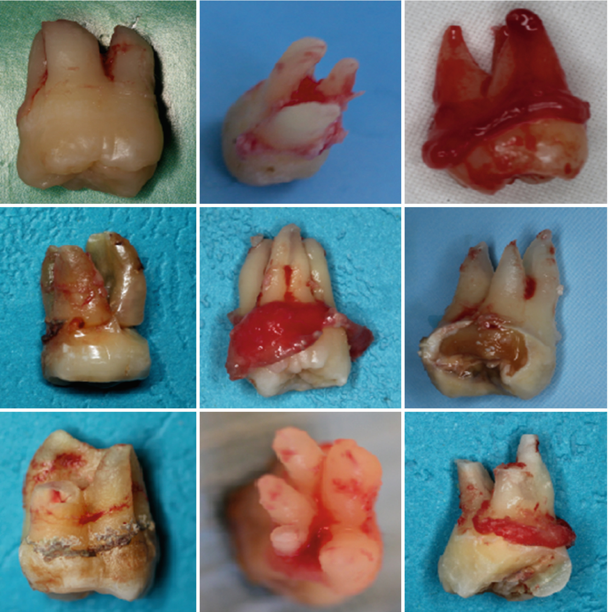
5 Roots In Molar
Wisdom tooth with 5 roots. One looks like a lil pecker! Posted by 5 years ago. Even as a dentist this is WTF. Wisdom tooth with 5 roots. A maxillary first molar has typically three separate roots and in only about 4% of the cases just two roots are found. Two or more merged roots occur in about 5% of all cases. The presence of four roots is extremely rare. In second maxillary molars, merging of roots is much more common.

- Maxillary First Molars - three well separated roots - palatal root - longest - mesio buccal root- broad bucco-lingually - mostly 2 canals - distobuccal - smallest root - straightest root - always look for four canals in all first molars - second mesial canal usually located in line with the groove between the mesiobuccal and palatal canals.
- If you have root decay, there is a high chance root canal treatment may be necessary to prevent the spread of decay and save your tooth. Root cavities are closer to the dental pulp in teeth, which means there is a higher chance bacteria will spread to the pulp.
SUMMARY
A total of 328 periapical x-rays from 105 patients of Mongolian origin, 106 of Negro origin and 117 of Caucasian origin were studied. The Mongolian race showed a greater incidence of three-rooted mandibular molars (15.2% of the Mongolian patients, 7.5% of the Negro patients and 6.8 of die Caucasian patients). There was no statistical difference in relation to sex and the incidence of this extra root.
Key Words: three-rooted mandibular molars, anatomy.
INTRODUCTION MATERIAL AND METHODS RESULTS DISCUSSION CONCLUSIONS REFERENCES
Introduction
The anatomy of human teeth present racial variations which can lead to therapy failure when not recognized. The failure of localization, instrumentation, and obturation of a root canal leads to problems which could be avoided.
Pucci and Reig (1944) verified an incidence of 5.5%of mandibular molars with 3 roots in a sample of teeth from the population of Uruguay. De Deus (1960) reports an incidence of 2.5% of these molars with 3 roots in a sample of teeth from patients in Southeastem Brazil. Teixeira (1963), citing an incidence of 10%, reported this extra root to be smaller than normal roots and in the disto-lingual position. Sousa-Freitas et al. (1971), using radiographic examinations, observed a presence of 17.8% of the mandibular first molars with 3 roots in patients of Japanese descent and of only 4.3% in patients of European descent. According to a review of the literature, a high incidence of mandibular molars with three roots is found in people of Mongolian origin (Japanese, Malaysian, Chinese, Thai, Eskimo, Aleutian, American Indian) (Tratman, 1938; Curzon, 1971; Jones, 1980; Reichart andMetah, 1981; Walkerand Quackenbush, 1985). The literature is lacking in studies about the incidence of this racial anatomic alteration in Brazil.
The objective of this research was to verify the incidence of three roots in human mandibular molars in patients of Mongolian, Caucasian (white) and Negro origin in the region of Ribeirão Preto, São Paulo, Brazil.
Material and Methods
A total of 328 periapical x-rays from 105 patients of Mongolian origin, 106 of Negro origin and 117 of Caucasian origin were analyzed. The molars were x-rayed by the long cone technique using a Dabi-Atlante (Ribeirão Preto, Brazil) x-ray machine with a 70-Kvp capacity. Kodak Ultraspeed films were used. For analysis of the x-rays, a negatoscope and a 4X lens were used. When the x-ray was not clear, a new one was taken changing the horizontal angle. Racial origin and sex were recorded.

Results
The presence of 3 roots in mandibular molars was confirmed in 16 patients of Mongolian origin (15.2%), in 8 patients of Negro origin (7.5%)and in 8 Caucasian patients (6.8%) (Table 1). There was a statistically significant difference (P < 0.01) in the incidence in the Mongolian race compared to the Negro and Caucasian races, which were statistically similar.
Table 1 - Mandibular molars with three roots fond III patients of Mongolian. Caucasian and Negro origin. The incidence of three-rooted mandibular molars in male and female patients is shown in table 2 according to racial origin. No significant statistical difference between males and females was found (Fisher test).
| Number of patiens | Race | Total | % | |||||||
| 105 | Mongolian | 12 (11,4%) | 3 (2,8%) | 1 (0,9%) | 16 | 15,2% | ||||
| 106 | Negro | 3 (2,8%) | 2 (1,8%) | 3 (2,8%) | 8 | 7,5% | ||||
| 117 | Caucasian | 5 (4,2%) | 2 (1,7%) | 1 (0,8%) | 8 | 6,8% | ||||
Table 2 - Incidence of mandibular molars with three roots according to race and sex.
| Total | |||
| Female | |||
| Mongolian | |||
| Negro | |||
| Caucasian | |||
| Total | |||
The incidence of first molars with three roots was 11.4% in patients of Mongolian origin, 2.8% in Negro patients and 4.2% in Caucasian patients.
The x-ray shown in Figure 1 is from a Caucasian patient whose mandibular first right molar had 3 roots and 4 root canals.
Discussion
The incidence of mandibular molars with three roots is high in people of Mongolian origin; however, it is also present in patients of Negro and Caucasian origin. this root is found in the disto-lingual position of mandibular molars.
Since the world today is no longer formed by races which do not mix, the dental surgeon must be aware of racial anatomical variations since he may see patients of diverse origins daily. In the region of Ribeirão Preto, Brazil, it is common to perform endodontic treatment on patients of Japanese, Chinese, Korean, White and Negro origin.
Table 3 shows the incidence of mandibular first molars with three roots in people of Mongolian origin reported in the literature. This table reports the possibility of these findings in a simple manner.
| Authors | Year | Origin | % |
| Tratman | 1938 | Malaysian | 12% |
| Tratman | 1938 | Chinese | 8% |
| Curzon | 1971 | Eskimo | 12,5% |
| Sousa-Freitas et al. | 1971 | Japanese descent | 22,7% |
| Somogyi | 1971 | American Indian | 16% |
| Jones | 1980 | Chinese | 13,4% |
| Jones | 1980 | Malaysian | 16% |
| Reichart and Metah | 1981 | Thai | 19,2% |
| Walker and Quackenbush | 1985 | Chinese 9Hong Kong) | 14,5% |
| Present study | 1992 | Japanese descent | 11,4% |
De Deus (1960) reported an incidence of mandibular first molars with 3 roots of only 2.5%, Teixeira (1963) reported 10% and Sousa-Freitas et al. (1971) observed 4.7%. We found an incidence of 4.2% in patients of Caucasian origin. In Negro patients, with an incidence of 7.5%of three-rooted mandibular molars, we found an incidence of 2.8% of first molars with 3 roots. the presence of 3-rooted mandibular molars is greater in patients of Mongolian origin but this does not lessen the importance of the occurrence in Negro and Caucasian patients.
It was not possible to verify the bilateral incidence since the patients studied lacked one or more mandibular molar.
Conclusions
1. the incidence of three-rooted mandibular molars is 15.2%in patients of Mongolian origin.
2. Negro patients presented an incidence of 3-rooted molars of 7.5%.
3. Caucasian patients (white) presented an incidence of 3-rooted mandibular molars of 6.8%, with 4.2% being first molars.
4. There was no statistical difference in the incidence of this dental anomaly in relation to sex.
References
Cruzon MEJ: Three-rooted mandibular permanent molars in the Keewatin Eskimo. Can Dent Assoc 37: 71-73, 1971
De Deus QD: Topografia da cavidade pulpar. Contribuição ao seu estudo. Doctorate thesis, Belo horizonte, 1960
Jones AW: The incidence of the three-rooted lower first permanent molar in malay people. Singapore Dent J 5:
15-17, 1980
Pucci FM, Reig R: Conductos Radiculares. Barreiro Y Ramos Montevideo, Vol 1, 1944
Reichart PA, Metah D: Three-rooted permanent mandibular first molars in flue Thai. Community Dent Oral Epidemiol 9: 191-192, 1981
Somogyi CW: Three-rooted mandibular first permanent molar in Alberta Indian children. Can Dent Assoc 37:105-106, 1971
Sousa-Freitas JA, Lopes ES, Casati-Alvares L: Anatomic variations of lower first permanent molar roots in two ethnic groups. Oral Surg 31: 274-278, 1971
Teixeira LD: Anatomia dentária humana. Imp Univ Minas Gerais, Belo Horizonte, 1963
Tratrnan EK: Three-rooted lower molars in man, and their racial distribution. Br Dent J 64: 264-267, 1938
Walker RT, Quackenbush LE: Three-rooted lower first permanent molars in Hong-Kong Chinese. Br Dent J 159:298-299, 1985
5 Roots In A Molar
Esta página foi elaborada com apoio do Programa Incentivo à Produção de Material Didático do SIAE - Pró - Reitorias de Graduação e Pós-Graduação da Universidade de São Paulo
Diagnostic:
D0120 Periodic exam: Periodic oral examination-established patient
D0140 Limited oral exam: Problem focused
D0150 Comprehensive oral exam: Extensive examination, new or established patient
D0160 Detailed and extensive oral evaluation: Problem focused, by report
D0170 Re-evaluation-limited, problem focused: Established patient, re-evaluation, not a post-op visit
D0171 Re-evaluation-post operative office visit: A recheck after a procedure to evaluate healing
D0460 Pulp vitality tests: Pulp testing
D0470 Diagnostic casts: Impressions and pouring up of plaster casts of teeth / dental arch
D9110 Emergency treatment: Palliative (emergency) pain relief – minor procedure
D9430 Office visit: Case follow-up/observation examination (during regular scheduled hours)
X-Rays:
D0210 Intraoral complete series of radiographic images: X rays of all teeth and the whole mouth
D0220 Intraoral periapical-first image: Detects changes/pathology @ root tip
D0230 Intraoral periapical-additional image(s)
D0270 Bitewing-single image: Detects changes/decay between teeth
D0272 Bitewing-two images
D0273 Bitewings-three images
D0274 Bitewings-four images
D0277 Vertical Bitewings: 7-8 bitewing images taken in the portrait orientation
D0330 Panoramic radiographic image: A 2-dimentional image of the whole mouth and teeth
D0364 Cone beam CT capture and interpretation limited view: Less than one whole jaw
D0365 Cone beam CT capture and interpretation limited view: Full lower jaw (mandible)
D0366 Cone beam CT capture and interpretation limited view: Full upper jaw(maxilla)
Interpretation and Report by a Practitioner Not Associated with the Capture: (D0380-D0391)
D0380 Cone beam CT interpretation limited view: Less than one whole jaw
D0381 Cone beam CT interpretation limited view: Full lower jaw (mandible)
D0382 Cone beam CT interpretation limited view: Full upper jaw (maxilla)
D0391 Interpretation of a diagnostic image by a practitioner
Tests and Examinations
D0460 Pulp vitality tests: Tests to determine which tooth (or teeth) are normal or diseased/need RCT or EXT
D0476 Special stains for microorganisms: Gram stains to determine the type of bacteria are present
Preventative
D1110 Prophylaxis-adult: Routine teeth cleaning/polish
D1120 Prophylaxis-child: Routine teeth cleaning/polish
D1206 Topical application of fluoride-varnish: A “paint-on” sticky fluoride application
D1208 Topical application of fluoride-excluding varnish: Gel fluoride application via trays
D1351 Sealant application-per tooth: A flowable acrylic that seals pits and fissures on teeth
D1353 Sealant repair-per tooth: Repair of a previous sealant that has worn or has debonded
Non-Surgical Endodontics:
D3120 Pulp cap (indirect): Removal of decay/site medication to heal pulp
D3220 Therapeutic pulpotomy: Emergency pulp chamber tissue removal for toothache relief
D3221 Gross pulpal debridement: Removal of complete pulpal tissue for toothache relief
D3230 Pulpal therapy (resorbable filling) anterior primary teeth: A root canal on a baby front tooth
D3240 Pulpal therapy (resorbable filling) posterior primary teeth: A root canal on a baby back tooth
D3310 Root canal-anterior: Root canal: front tooth
D3320 Root canal-bicuspid: Root canal: middle tooth
D3330 Root canal-molar: Root canal: back tooth
D3331 Treatment of root canal obstruction: Removal of a separated instrument, finding a Ca+ canal
D3332 Incomplete root canal therapy: Inoperable or fractured tooth, root canal procedure not completed
D3333 Internal root repair: Repair of perforation defects
D3346 Retreatment-anterior: To re-do a failing root canal: front tooth
D3347 Retreatment-bicuspid: To re-do a failing root canal: middle tooth
D3348 Retreatment-molar: To re-do a failing root canal: back tooth
D3351 Apexification-initial: To close/complete root tip development: Visit #1
D3552 Apexification-interim: Multiple visits/dressing change: Visit #2+
5 Roots In Molar Formula
D3553 Apexification-final: Completion of root apex closure: Final visit
D3351 Calcification/repair: To induce bone growth to seal root surfaces (i.e. perforations, resorption)
D3355 Pulpal regeneration: Procedures to induce regrowth of pulpal tissue in an immature tooth
D3356 Pulpal regeneration-interim visit: Change of medication
D3357 Pulpal regeneration-completion of treatment: Removal of canal medication and filling of the canal
Endodontic Surgery:
D3410 Apicoectomy-anterior: Root tip surgery: front tooth
D3421 Apicoectomy-bicuspid: Root tip surgery: middle tooth
D3425 Apicoectomy-molar: Root tip surgery:back tooth
D3426 Apicoectomy-additional root: Root tip surgery: extra roots
D3428 Bone graft in conjunction with periapical surgery: Placement of biologics to aid in healing
D3429 Bone graft in conjunction with periapical surgery: Each additional tooth in the same site
D3430 Retrograde filling: A surgically placed root-end filling
D3431 Biologic materials to aid in bone and soft tissue healing
D3432 Guided tissue regeneration: Placement of a resorbable barrier to aid in root surgery
D3450 Root amputation: Removal of one root of a tooth
D3470 Intentional reimplantation: Removing/treating/replacing a tooth into its own socket
D3910 Surgical procedure for the isolation of a tooth: Procedures to help attach a rubber dam
D3920 Hemisection: Sectioning/removal of half of two rooted tooth
D4249 Crown lengthening: Procedures to enhance/expose root for restorative enhancement
D0501 Histopathologic Exam: Sampling and microscopic examination of oral disease
D7111 Extraction of a primary tooth: Removal of the remnants of a “baby” tooth
D7140 Extraction of a permanent tooth: Forceps removal of an “adult” tooth
D7210 Extraction of a permanent tooth: Removal of an adult tooth requiring flap, bone removal
D7270 Reimplantation/splint: Replacing/stabilizing a knocked-out tooth
D7272 Tooth transplantation: Extraction and moving/implanting the tooth to a different site in the mouth
D7285 Biopsy-hard tissue: Sampling and microscopic examination of bone or tooth structure
D7286 Biopsy-soft tissue: Sampling and microscopic examination of surgical soft tissue
D7288 Brush biopsy: Sampling of oral soft tissue using a brush for microscopic examination
D7430 Cystectomy (<1.25cm): Removal of a small cyst
D7431 Cystectomy (>1.25cm): Removal of a large cyst
D7510 Incision and drainage: Lancing a swollen area to relieve infection
D7270 Reimplantation/splint: Replacing/stabilizing a knocked-out tooth
Restorative:
D2140 to 61 Amalgam: Placement of a metal, compactable filling, 1 to 4 surfaces.
5 Roots In Molars
D2330 to 94 Composite: Placement/bonding of a plastic tooth colored filling, 1 to 4 surfaces
D2920 Re-cement crown: Reglue a single crown to tooth
D2952 Cast gold post/core: A casted single unit post/core to attach a crown to a root
D2954 Prefabricated post/core: A standard post/core to attach a crown to a root
D2955 Post removal: Using ultrasonics and operating microscope to remove a post
D2970 Temporary crown: To make a temporary crown or “cap”
D2980 Crown repair: To repair a crown necessitated by restorative material failure
D3950 Post space preparation: Removal of a root canal filling for a post
D6930 Re-cement bridge: Reglue multiple, attached crowns to teeth
D9120 Sectioning of a fixed bridge: Cutting apart and removing part of a bridge
Drugs:
D9610 Therapeutic drug: By injection, single administration, one medication
D9612 Therapeutic drugs: By injection, two or more administrations, different medications
Other:
D3960 Internal bleaching initial/subsequent visits: To lighten a single, dark tooth
D3999 Unspecified: Misc., by report
D5410 Adjustment of a complete denture-maxillary (“upper”)
D5411 Adjustment of a complete denture-mandibular (“lower”)
D5421 Adjustment of a partial denture-maxillary (“upper”)

D5422 Adjustment of a partial denture-mandibular (“lower”)
D9910 Desensitizer application: Medication to decrease thermal pain
D9943 Occlusal guard adjustment: Adjustment of a night guard or bruxing splint
D9951 Occlusal adjustment: Selective bite adjustment
Many of the listed procedures do not fall under what is considered “mainstream endodontics” as a specialty, but include procedures typically offered in a general practice or other dental specialties like oral surgery, periodontics, prosthodontics, etc. They are listed because of our experience that many of the patients that we treated under moderate to deep sedation/general anesthesia needed the other non-endodontic procedures performed while they were asleep. Completing these procedures in conjunction with the endodontic treatment would enhance the success of the case, help get the patient out of pain, or prevent another problem “waiting to happen” while the patient was under anesthesia just the one time.

5 Roots In Molar Volume
This saves the patient time, money, minimizes risk and promotes a culture of safety.